How to Read Ct Sinus Scans Like an Expert
Computed Tomography (CT) - Sinuses
Computed tomography (CT) of the sinuses uses special x-ray equipment to evaluate the paranasal sinus cavities – hollow, air-filled spaces within the bones of the face surrounding the nasal cavity. CT scanning is painless, noninvasive and accurate. It'south also the near reliable imaging technique for determining if the sinuses are obstructed and the best imaging modality for sinusitis.
Tell your doctor if in that location's a possibility y'all are significant and discuss any recent illnesses, medical conditions, medications you're taking, and allergies. This exam does non usually require dissimilarity material; however, in some situations your doctor may asking that contrast material exist given. If y'all have a known allergy to dissimilarity material, your doctor may prescribe medications to reduce the risk of an allergic reaction. These medications must exist taken 12 hours prior to your exam. Get out jewelry at home and wear loose, comfy clothing. Y'all may need to change into a gown for the procedure.
- What is CT (Computed Tomography) of the Sinuses?
- What are some common uses of the procedure?
- How should I prepare?
- What does the equipment expect like?
- How does the procedure piece of work?
- How is the procedure performed?
- What volition I feel during and after the process?
- Who interprets the results and how practice I go them?
- What are the benefits vs. risks?
- What are the limitations of CT of the Sinuses?
- Which exam, process or treatment is best for me?
What is CT (Computed Tomography) of the Sinuses?
Computed tomography, more commonly known every bit a CT or CAT scan, is a diagnostic medical imaging test. Similar traditional x-rays, it produces multiple images or pictures of the inside of the body.
A CT scan generates images that can be reformatted in multiple planes. It tin even generate 3-dimensional images. Your doctor tin can review these images on a computer monitor, print them on film or via a 3D printer, or transfer them to a CD or DVD.
CT images of internal organs, basic, soft tissue, and blood vessels provide greater particular than traditional x-rays. This is especially truthful for soft tissues and blood vessels.
A CT browse of the face produces images that also show a patient's paranasal sinus cavities. The paranasal sinuses are hollow, air-filled spaces located within the bones of the face and surrounding the nasal cavity, a organization of air channels connecting the nose with the back of the throat. In that location are iv pairs of sinuses, each connected to the nasal crenel by small openings.
top of page
What are some mutual uses of the procedure?
CT of the sinuses is primarily used to:
- help diagnose sinusitis.
- evaluate sinuses that are filled with fluid or thickened sinus membranes.
- observe the presence of inflammatory diseases.
- provide additional information about tumors of the nasal crenel and sinuses.
- plan for surgery past defining anatomy.
top of page
How should I set up?
Vesture comfortable, loose-plumbing fixtures clothing to your exam. You may need to modify into a gown for the process.
Metal objects, including jewelry, eyeglasses, dentures, and hairpins, may bear on the CT images. Leave them at home or remove them prior to your examination. Some CT exams will crave you to remove hearing aids and removable dental piece of work. Women will need to remove bras containing metallic underwire. Yous may need to remove any piercings, if possible.
Your doctor may instruct you to not eat or drinkable anything for a few hours earlier your exam if it will use dissimilarity textile. Tell your doctor virtually all medications you are taking and if yous have whatsoever allergies. If you have a known allergy to contrast material, your doc may prescribe medications (commonly a steroid) to reduce the risk of an allergic reaction. To avoid unnecessary delays, contact your doctor well earlier the date of your exam.
Also tell your doctor about whatever contempo illnesses or other medical conditions and whether you lot take a history of heart disease, asthma, diabetes, kidney disease, or thyroid problems. Any of these conditions may increase the risk of an agin consequence.
Women should always inform their physician and the CT technologist if there is any possibility that they may be meaning. Run into the CT Safety During Pregnancy page for more than information.
top of page
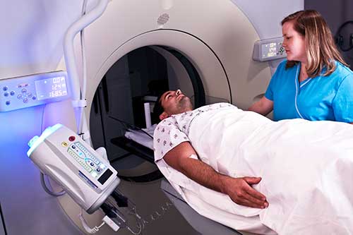
What does the equipment look like?
The CT scanner is typically a big, donut-shaped machine with a curt tunnel in the center. You volition lie on a narrow table that slides in and out of this short tunnel. Rotating effectually you, the x-ray tube and electronic 10-ray detectors are located opposite each other in a ring, called a gantry. The computer workstation that processes the imaging information is in a separate control room. This is where the technologist operates the scanner and monitors your test in directly visual contact. The technologist will exist able to hear and talk to you using a speaker and microphone.
top of folio
How does the procedure piece of work?
In many ways, a CT scan works like other ten-ray exams. Different body parts absorb ten-rays in different amounts. This difference allows the doctor to distinguish body parts from one some other on an x-ray or CT image.
A conventional ten-ray examination directs a minor amount of radiation through the body part nether examination. A special electronic image recording plate captures the image. Bones appear white on the x-ray. Soft tissue, such as the center or liver, shows up in shades of gray. Air appears black.
With CT scanning, several ten-ray beams and electronic x-ray detectors rotate around y'all. These measure out the amount of radiations being captivated throughout your trunk. Sometimes, the exam table will move during the browse. A special calculator program processes this big volume of information to create ii-dimensional cross-sectional images of your trunk. The system displays the images on a figurer monitor. CT imaging is sometimes compared to looking into a loaf of bread past cutting the loaf into sparse slices. When the computer software reassembles the epitome slices, the upshot is a very detailed multidimensional view of the body's interior.
Nearly all CT scanners tin obtain multiple slices in a unmarried rotation. These multi-slice (multidetector) CT scanners obtain thinner slices in less time. This results in more detail.
Modern CT scanners can image big sections of the trunk in just a few seconds, and even faster in small children. Such speed is benign for all patients. Speed is particularly benign for children, the elderly, and critically ill – anyone who finds it hard to stay still, even for the brief time necessary to obtain images.
For children, the radiologist will adjust the CT scanner technique to their size and the surface area of interest to reduce the radiation dose.
Some CT exams use a dissimilarity material to enhance visibility in the body expanse under examination.
peak of page
How is the procedure performed?
The technologist begins by positioning patients on the CT examination table.
For a CT scan of the sinuses, the patient is about commonly positioned lying flat on the back. The patient may also be positioned face-downward with the chin elevated.
Straps and pillows may be used to assistance the patient maintain the right position and to hold still during the examination.
Some patients require an injection of a contrast material to enhance the visibility of certain tissues or blood vessels. If contrast material is required, a nurse or technologist will insert an intravenous (IV) line into a small-scale vein in the patient'due south hand or arm. The dissimilarity cloth will exist injected through this line.
Side by side, the table volition move quickly through the scanner to determine the correct starting position for the scans. And then, the table will move slowly through the machine for the actual CT scan. Depending on the type of CT browse, the machine may make several passes.
The technologist may ask you to concord your jiff during the scanning. Any motion, including breathing and torso movements, can lead to artifacts on the images. This loss of image quality can resemble the blurring seen on a photograph taken of a moving object.
When the test is complete, the technologist will ask you lot to wait until they verify that the images are of loftier plenty quality for authentic interpretation by the radiologist.
The actual CT scan takes less than a minute and the entire process is usually completed inside 10 minutes.
top of page
What will I experience during and afterward the procedure?
CT exams are mostly painless, fast, and easy. Multidetector CT reduces the amount of fourth dimension that the patient needs to prevarication still.
Though the scanning itself causes no pain, at that place may exist some discomfort from having to remain nonetheless for several minutes. If you have a hard time staying still, are claustrophobic, or accept chronic pain, y'all may find a CT exam to be stressful. The technologist or nurse, under the management of a physician, may offer you some medication to help you lot tolerate the CT scanning procedure.
If the exam uses iodinated contrast material, your doctor will screen you for chronic or acute kidney disease. The doctor may administer dissimilarity material intravenously (past vein), then you will feel a pin prick when the nurse inserts the needle into your vein. You lot may feel warm or flushed as the contrast is injected. You besides may have a metal sense of taste in your rima oris. This will laissez passer. You may feel a need to urinate. Yet, these are only side effects of the contrast injection, and they subside quickly.
When you enter the CT scanner, you may run into special light lines projected onto your body. These lines help ensure that y'all are in the correct position on the exam table. With modern CT scanners, you may hear slight buzzing, clicking and whirring sounds. These occur equally the CT scanner's internal parts, not usually visible to you, circumduct around you lot during the imaging procedure.
You will be lone in the test room during the CT scan, unless in that location are special circumstances. For example, sometimes a parent wearing a lead shield may stay in the room with their child. Nevertheless, the technologist will e'er be able to see, hear and speak with you through a built-in intercom system.
With pediatric patients, a parent may be allowed in the room but may demand to article of clothing a lead apron to minimize radiations exposure.
After a CT exam, the technologist will remove your intravenous line. They will comprehend the tiny hole made past the needle with a small-scale dressing. You can return to your normal activities immediately.
top of page
Who interprets the results and how do I get them?
A radiologist, a md specially trained to supervise and interpret radiology exams, will analyze the images. The radiologist will transport an official study to the doctor who ordered the examination.
You may need a follow-up test. If so, your doc will explain why. Sometimes a follow-upwardly test further evaluates a potential issue with more views or a special imaging technique. It may besides come across if there has been any change in an result over time. Follow-up exams are often the best way to run across if handling is working or if a trouble needs attending.
top of page
What are the benefits vs. risks?
Benefits
- CT scan is ane of the safest means of studying the sinuses.
- CT is the nearly reliable imaging technique for determining if the sinuses are obstructed. It is the all-time imaging modality for sinusitis.
- CT of the sinuses can help plan the safest and about effective surgery.
- CT of the sinuses is now widely available and is performed in a relatively short time, especially when compared to magnetic resonance imaging (MRI).
- CT scanning is painless, noninvasive, and accurate.
- A major advantage of CT is its power to prototype os, soft tissue, and blood vessels all at the same time.
- Dissimilar conventional x-rays, CT scanning provides very detailed images of many types of tissue too every bit the lungs, bones, and blood vessels.
- CT exams are fast and simple. In emergency cases, they can reveal internal injuries and haemorrhage quickly plenty to aid salvage lives.
- CT has been shown to be a toll-effective imaging tool for a wide range of clinical issues.
- CT is less sensitive to patient movement than MRI.
- Unlike MRI, an implanted medical device of any kind volition non foreclose you lot from having a CT browse.
- CT imaging provides real-time imaging, making it a good tool for guiding needle biopsies and needle aspirations. This is peculiarly true of procedures involving the lungs, abdomen, pelvis, and bones.
- A diagnosis via CT scan may eliminate the need for exploratory surgery and surgical biopsy.
- No radiation remains in a patient's body after a CT exam.
- The x-rays used for CT scanning should have no firsthand side effects.
Risks
- There is always a slight gamble of cancer from excessive exposure to radiation. However, the benefit of an authentic diagnosis far outweighs the risk involved with CT scanning.
- The radiation dose for this procedure varies. See the Radiation Dose in X-Ray and CT Exams folio for more information virtually radiation dose.
- Women should always tell their medico and x-ray or CT technologist if there is whatever risk they are pregnant. See the Safety in X-ray, Interventional Radiology and Nuclear Medicine Procedures page for more information about pregnancy and x-rays.
- Doctors exercise not generally recommend CT scanning for pregnant women unless medically necessary because of potential risk to the unborn babe.
- IV dissimilarity manufacturers betoken mothers should non breastfeed their babies for 24-48 hours after contrast material is given. However, the most recent American College of Radiology (ACR) Transmission on Contrast Media reports that studies show the amount of contrast absorbed by the infant during breastfeeding is extremely low. For further information please consult the ACR Manual on Contrast Media and its references.
- The hazard of serious allergic reaction to dissimilarity materials that contain iodine is extremely rare, and radiology departments are well-equipped to deal with them.
- Because children are more sensitive to radiations, they should have a CT exam only if it is essential for making a diagnosis. They should not accept repeated CT exams unless necessary. CT scans in children should always exist done with low-dose technique.
top of page
What are the limitations of CT of the Sinuses?
CT is usually the first exam ordered when a sinus tumor is suspected. If additional data is needed to determine the extent of soft tissue of the tumor, magnetic resonance imaging (MRI) may be helpful.
A person who is very large may not fit into the opening of a conventional CT scanner. Or, they may be over the weight limit—usually 450 pounds—for the moving table.
top of page
pinnacle of page
This page was reviewed on June, 15, 2020
Images
Pediatric Content
Some imaging tests and treatments take special pediatric considerations. The teddy bear denotes child-specific content.
- RadInfo 4 Kids
Sponsored Past
Please notation
RadiologyInfo.org is not a medical facility. Delight contact your medico with specific medical questions or for a referral to a radiologist or other physician. To locate a medical imaging or radiation oncology provider in your community, yous tin search the ACR-accredited facilities database.
This website does not provide toll information. The costs for specific medical imaging tests, treatments and procedures may vary by geographic region. Discuss the fees associated with your prescribed procedure with your dr., the medical facility staff and/or your insurance provider to go a better understanding of the possible charges you will incur.
Web page review procedure: This Spider web page is reviewed regularly by a physician with expertise in the medical expanse presented and is further reviewed by committees from the Radiological Society of North America (RSNA) and the American College of Radiology (ACR), comprising physicians with expertise in several radiologic areas.
Outside links: For the convenience of our users, RadiologyInfo.org provides links to relevant websites. RadiologyInfo.org, RSNA and ACR are not responsible for the content contained on the web pages constitute at these links.
Source: https://www.radiologyinfo.org/en/info/sinusct
0 Response to "How to Read Ct Sinus Scans Like an Expert"
Post a Comment